In An Ecg Pattern The P Wave Is Caused By
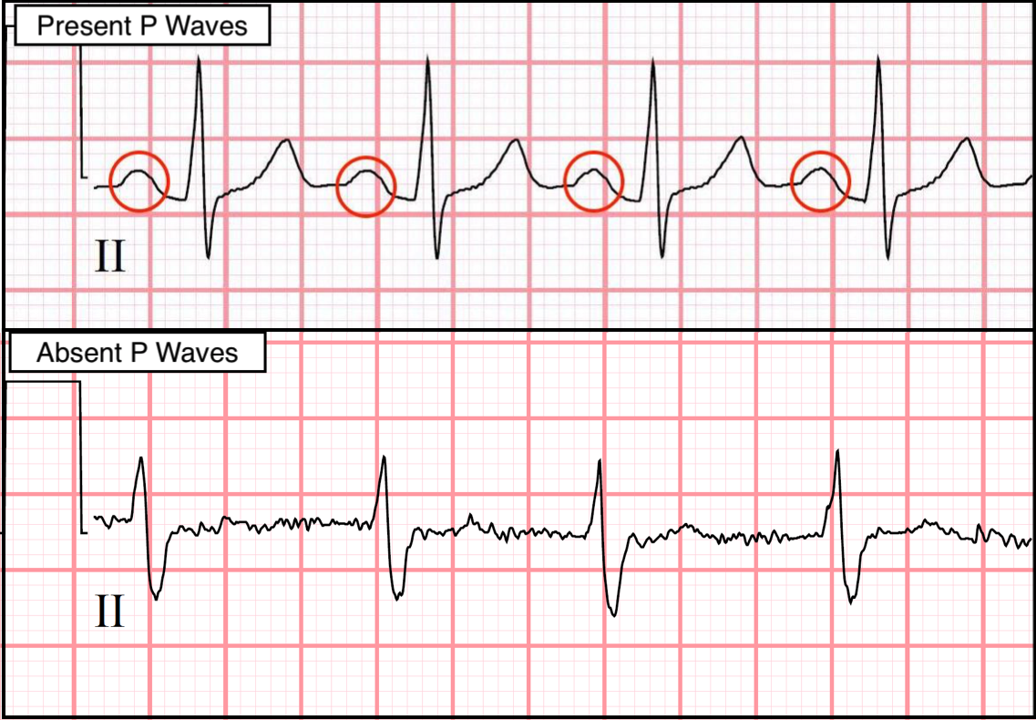
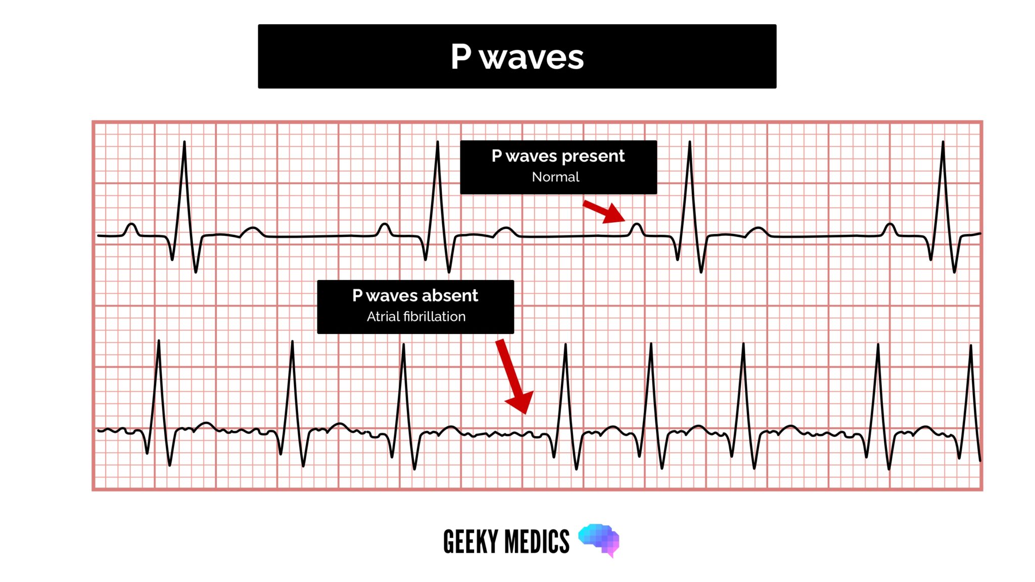
In An Ecg Pattern The P Wave Is Caused By - Web inverted p wave (ecg) an inverted p wave on an ecg is usually a sign of ectopic atrial rhythm. It represents the electrical activity associated with atrial depolarization. Web parts of the ecg explained p waves. Web a premature atrial complex (pac) is a premature beat arising from ectopic pacemaking tissue within the atria. The p wave is a summation wave generated by the depolarization front as it transits the atria. Web the factors that determine p‐wave appearance include (1) the origin of the sinus rhythm that defines right atrial depolarization vector, (2) localization of left atrial. The right atrium (ra) is depolarized towards the av node. The normal p wave is a low. Web learn the heart is a comprehensive guide to understanding p waves and their significance in ecg interpretation. Web the p wave represents the time during which the electrical impulse travels through the atria, causing depolarization and leading to their contraction. There is an abnormal p wave, usually followed by a. P waves represent atrial depolarisation. Web learn the heart is a comprehensive guide to understanding p waves and their significance in ecg interpretation. Normally the right atrium depolarizes slightly earlier than left atrium since the depolarization wave originates in the sinoatrial node, in the high right atrium and then travels to and through the left atrium. The key points on those waves are labeled p, q, r, s, and t. Web chronic pulmonary hypertension leading to chronic right atrial and ventricular hypertrophy and dilation may manifest as p waves of higher amplitude (p pulmonale). Web the factors that determine p‐wave appearance include (1) the origin of the sinus rhythm that defines right atrial depolarization vector, (2) localization of left atrial. Web the p wave represents the time during which the electrical impulse travels through the atria, causing depolarization and leading to their contraction. Web inverted p wave (ecg) an inverted p wave on an ecg is usually a sign of ectopic atrial rhythm. Web the p wave is the first wave on the ecg because the action potential for the heart is generated in the sinoatrial (sa) node, located on the atria, which sends action. Web a premature atrial complex (pac) is a premature beat arising from ectopic pacemaking tissue within the atria. Web inverted p wave (ecg) an inverted p wave on an ecg is usually a sign of ectopic atrial rhythm. The normal p wave is a low. There is an abnormal p wave, usually followed by a. Web the p wave is. The action potentials that initiate myocardiocyte depolarization. The key points on those waves are labeled p, q, r, s, and t. Web the p wave is the first wave on the ecg because the action potential for the heart is generated in the sinoatrial (sa) node, located on the atria, which sends action. Web the p wave is the first. Web chronic pulmonary hypertension leading to chronic right atrial and ventricular hypertrophy and dilation may manifest as p waves of higher amplitude (p pulmonale). Normally the right atrium depolarizes slightly earlier than left atrium since the depolarization wave originates in the sinoatrial node, in the high right atrium and then travels to and through the left atrium. The normal p. It represents the electrical activity associated with atrial depolarization. Web the p wave represents atrial depolarization, which starts in the sinus node. The normal p wave is a low. Web the p wave is the first deflection of the cardiac cycle seen on an ekg. Web your solution’s ready to go! Web your solution’s ready to go! Web learn the heart is a comprehensive guide to understanding p waves and their significance in ecg interpretation. It represents the electrical activity associated with atrial depolarization. Web a premature atrial complex (pac) is a premature beat arising from ectopic pacemaking tissue within the atria. The key points on those waves are labeled p,. Web the p wave morphology can reveal right or left atrial hypertrophy or atrial arrhythmias and is best determined in leads ii and v1 during sinus rhythm. Web the factors that determine p‐wave appearance include (1) the origin of the sinus rhythm that defines right atrial depolarization vector, (2) localization of left atrial. Web parts of the ecg explained p. The action potentials that initiate myocardiocyte depolarization. Web the p wave is the first deflection of the cardiac cycle seen on an ekg. Web learn the heart is a comprehensive guide to understanding p waves and their significance in ecg interpretation. It represents the electrical activity associated with atrial depolarization. Web the p wave morphology can reveal right or left. In healthy individuals, there should be a p wave preceding each qrs. Web learn the heart is a comprehensive guide to understanding p waves and their significance in ecg interpretation. Web the p wave is the first wave on the ecg because the action potential for the heart is generated in the sinoatrial (sa) node, located on the atria, which. Web in an ecg pattern, the pq interval indicates how long it takes the cardiac impulse ti travel from the The key points on those waves are labeled p, q, r, s, and t. Web chronic pulmonary hypertension leading to chronic right atrial and ventricular hypertrophy and dilation may manifest as p waves of higher amplitude (p pulmonale). Web parts. Web inverted p wave (ecg) an inverted p wave on an ecg is usually a sign of ectopic atrial rhythm. Web an ecg readout represents the pattern of electrical activity in the heart as a line of waves. The key points on those waves are labeled p, q, r, s, and t. Question 42 in an ecg pattern,. Web the. P waves represent atrial depolarisation. The key points on those waves are labeled p, q, r, s, and t. Web the p wave is the first deflection of the cardiac cycle seen on an ekg. In healthy individuals, there should be a p wave preceding each qrs. The normal p wave is a low. Web a premature atrial complex (pac) is a premature beat arising from ectopic pacemaking tissue within the atria. Web the p wave is the first wave on the ecg because the action potential for the heart is generated in the sinoatrial (sa) node, located on the atria, which sends action. Question 42 in an ecg pattern,. Web in an ecg pattern, the pq interval indicates how long it takes the cardiac impulse ti travel from the Web the p wave represents atrial depolarization, which starts in the sinus node. Web your solution’s ready to go! Web the p wave represents the time during which the electrical impulse travels through the atria, causing depolarization and leading to their contraction. Normally the right atrium depolarizes slightly earlier than left atrium since the depolarization wave originates in the sinoatrial node, in the high right atrium and then travels to and through the left atrium. Web the factors that determine p‐wave appearance include (1) the origin of the sinus rhythm that defines right atrial depolarization vector, (2) localization of left atrial. The p wave is a summation wave generated by the depolarization front as it transits the atria. There is an abnormal p wave, usually followed by a.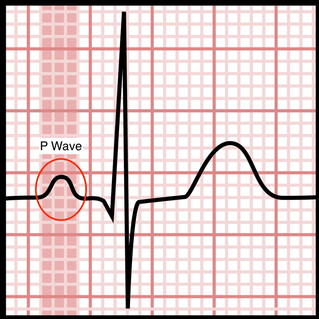
Electrocardiograms (EKGs/ECGs) Evaluating P Waves Stepwards
ECG Waveform Explained EKG Labeled Diagrams and Components — EZmed

Pwave, PR interval, PR segment physiology, criteria & ECG findings
Electrocardiograms (EKGs/ECGs) Evaluating P Waves Stepwards
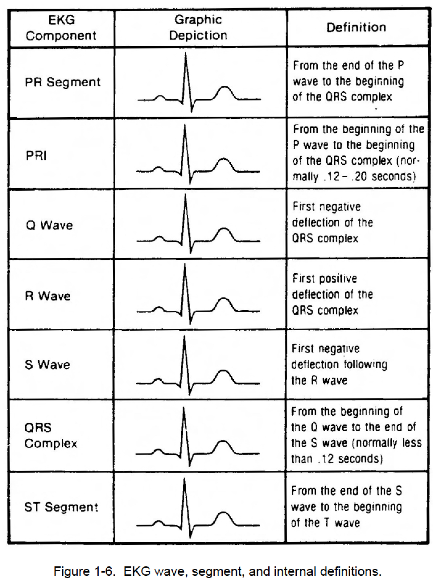
Figure 16. EKG wave, segment, and internal definitions Cardiac
Electrocardiograms (EKGs/ECGs) Evaluating P Waves Stepwards
How to Read an ECG ECG Interpretation EKG Geeky Medics

P wave wikidoc

Pwave, PR interval, PR segment physiology, criteria & ECG findings

ECG interpretation Characteristics of the normal ECG (Pwave, QRS
It Represents The Electrical Activity Associated With Atrial Depolarization.
The Pr Interval Is Assessed In Order To Determine Whether Impulse Conduction From The Atria To The.
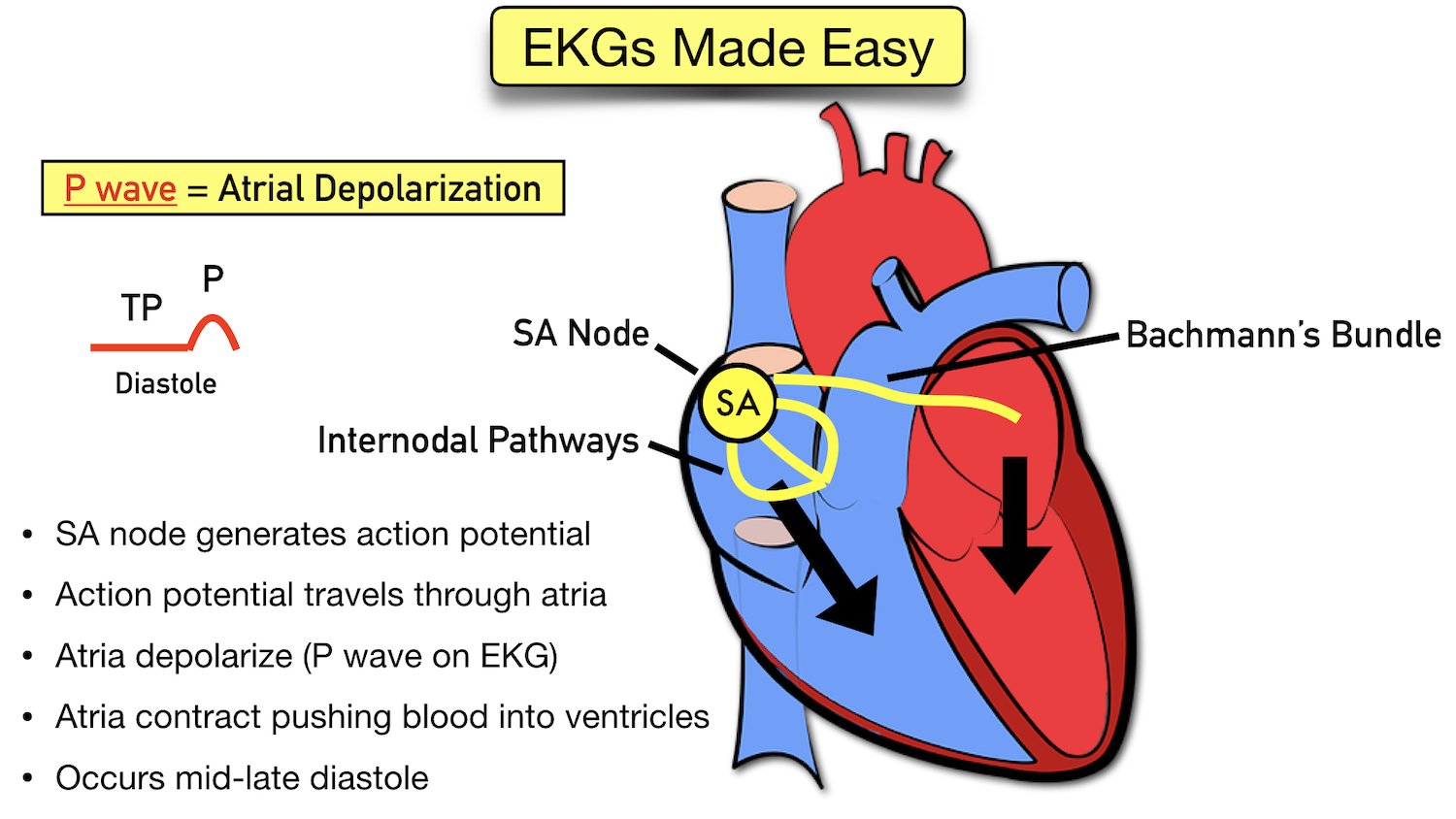
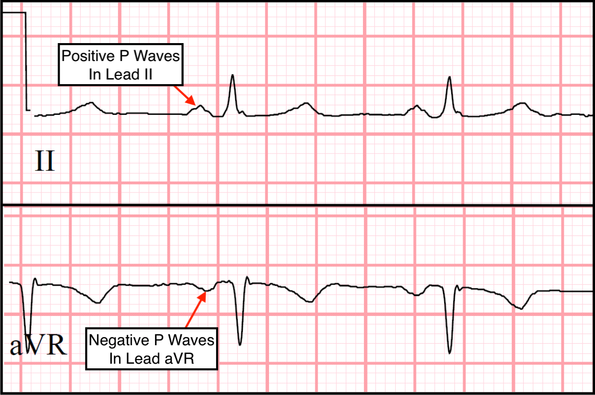
The Right Atrium (Ra) Is Depolarized Towards The Av Node.
Web Parts Of The Ecg Explained P Waves.
Related Post: