Lvh Strain Pattern
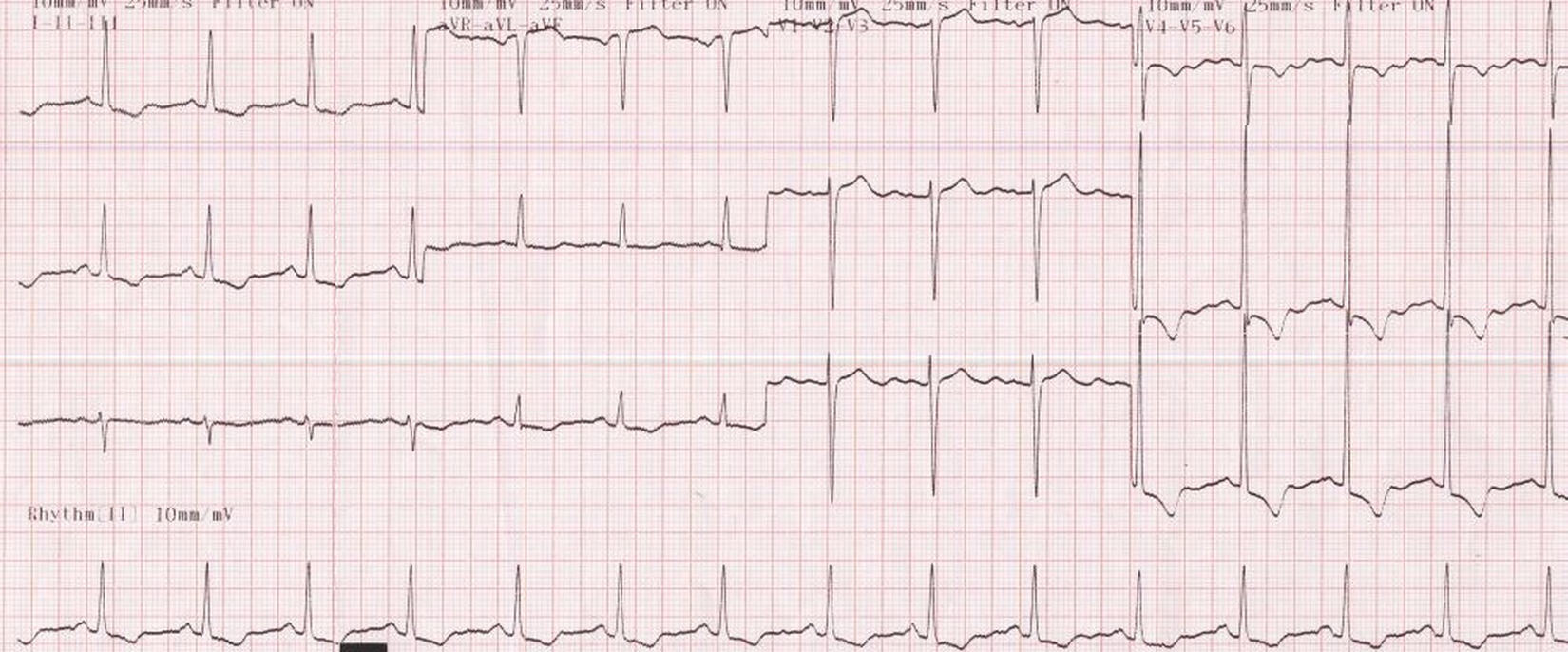
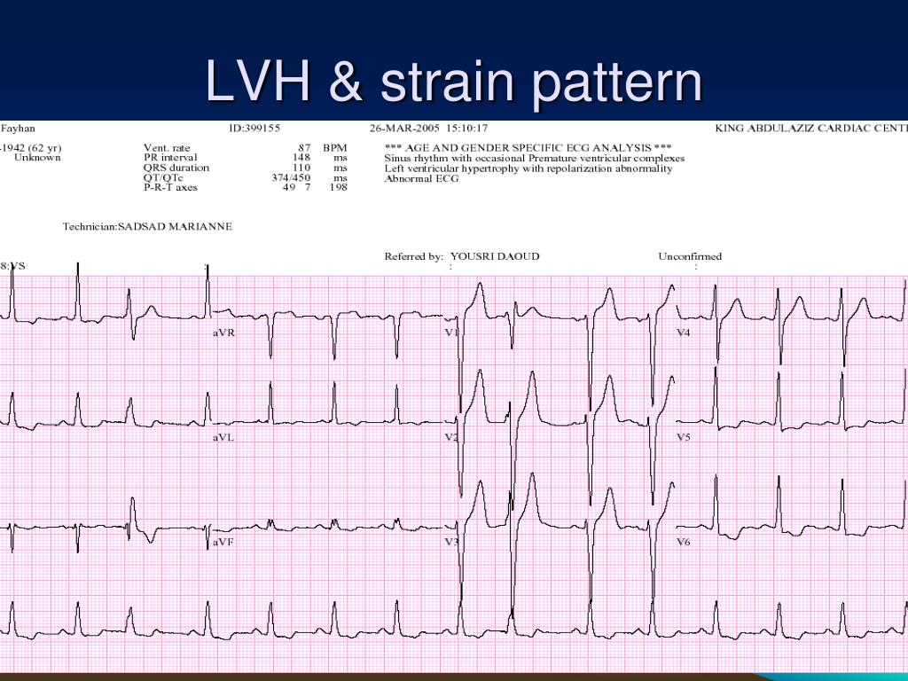
Lvh Strain Pattern - As both lvh and ivcds alter qrs patterns, the existence of an ivcd may impact the accuracy of ecg criteria for lvh. Such hypertrophy is usually the response to a chronic pressure or volume load. Web lvh with strain pattern can sometimes be seen in long standing severe aortic regurgitation, usually with associated left ventricular hypertrophy and systolic dysfunction. Web the most common causes of left ventricular hypertrophy are aortic stenosis, aortic regurgitation, hypertension, cardiomyopathy and coarctation of the aorta. Web patterns of lv geometry were defined according to lvh and rwt: Left ventricular hypertrophy itself doesn't cause symptoms. This is seen in sloping st depressions in all leads with upright qrs complexes. There will also be slight st elevations (reciprocal to the depressions) in leads with negative qrss. It can also cause changes to the heart’s conduction system that make it beat irregularly (arrhythmia). Web left ventricular hypertrophy (lvh) refers to an increase in the size of myocardial fibers in the main cardiac pumping chamber. The sensitivity of lvh strain pattern on ecg as a measure of lvh has ranged from 3.8% to 50% in various reports [1]. Some people do not have symptoms, especially during the early stages of the condition. Such hypertrophy is usually the response to a chronic pressure or volume load. Web patterns of lv geometry were defined according to lvh and rwt: Web the st changes in lvh are due to the strain pattern, indicating strain on the left ventricular myocardium. There will also be slight st elevations (reciprocal to the depressions) in leads with negative qrss. Web when lvh is caused by a pathological condition, we often see the strain pattern, which is st depression and t wave inversion in leads with upright qrs complexes (the lateral leads). It's important to manage conditions such as high blood pressure and sleep apnea, which can. As both lvh and ivcds alter qrs patterns, the existence of an ivcd may impact the accuracy of ecg criteria for lvh. It is true that some st elevation will appear in v1 and v2 in these patients, and can be mistaken for m.i. Web in order to diagnose lvh from the ecg, we must also show repolarization abnormalities, called the strain pattern. Web the most common causes of left ventricular hypertrophy are aortic stenosis, aortic regurgitation, hypertension, cardiomyopathy and coarctation of the aorta. It can also cause changes to the heart’s conduction system that make it beat irregularly (arrhythmia). However, whether ecg strain. It can also cause changes to the heart’s conduction system that make it beat irregularly (arrhythmia). Web left ventricular hypertrophy with strain pattern (example 3) | learn the heart. Web left ventricular hypertrophy commonly occurs in heart diseases that also cause intraventricular conduction defects or delays (ivcds). We investigated the mechanisms and outcomes associated with ecg strain. Web lvh with. However, whether ecg strain is an independent predictor of cardiovascular (cv) morbidity and mortality in the setting of aggressive antihypertensive therapy is unclear. There are several ecg indexes, which generally have high diagnostic specificity but low sensitivity. (1) normal (no lvh, normal rwt); As both lvh and ivcds alter qrs patterns, the existence of an ivcd may impact the accuracy. But symptoms may occur as the strain on the heart worsens. Web lvh with strain pattern can sometimes be seen in long standing severe aortic regurgitation, usually with associated left ventricular hypertrophy and systolic dysfunction. Left ventricular hypertrophy itself doesn't cause symptoms. Ecg‐lvh was assessed by qrs voltage and duration. Web the st changes in lvh are due to the. Web when lvh is caused by a pathological condition, we often see the strain pattern, which is st depression and t wave inversion in leads with upright qrs complexes (the lateral leads). The sensitivity of lvh strain pattern on ecg as a measure of lvh has ranged from 3.8% to 50% in various reports [1]. Such hypertrophy is usually the. Web the most common causes of left ventricular hypertrophy are aortic stenosis, aortic regurgitation, hypertension, cardiomyopathy and coarctation of the aorta. Web left ventricular hypertrophy (lvh) refers to an increase in the size of myocardial fibers in the main cardiac pumping chamber. Huge precordial r and s waves that overlap with the adjacent leads (sv2 + rv6 >> 35 mm).. Web left ventricular hypertrophy (lvh) refers to an increase in the size of myocardial fibers in the main cardiac pumping chamber. There are several ecg indexes, which generally have high diagnostic specificity but low sensitivity. (2) concentric remodeling (no lvh, increased rwt); Some people do not have symptoms, especially during the early stages of the condition. Note that the y. It may include medications, catheter procedures or surgery. We investigated the mechanisms and outcomes associated with ecg strain. Huge precordial r and s waves that overlap with the adjacent leads (sv2 + rv6 >> 35 mm). Note that the y ‐axis scales vary by outcome. There will also be slight st elevations (reciprocal to the depressions) in leads with negative. Web left ventricular hypertrophy with strain pattern ecg (example 1) | learn the heart. As both lvh and ivcds alter qrs patterns, the existence of an ivcd may impact the accuracy of ecg criteria for lvh. It is true that some st elevation will appear in v1 and v2 in these patients, and can be mistaken for m.i. 2,6 ecg. Some people do not have symptoms, especially during the early stages of the condition. Note that the y ‐axis scales vary by outcome. Web lvh with strain pattern can sometimes be seen in long standing severe aortic regurgitation, usually with associated left ventricular hypertrophy and systolic dysfunction. We investigated the mechanisms and outcomes associated with ecg strain. It may include. Web left ventricular hypertrophy with strain pattern (example 3) | learn the heart. (3) eccentric hypertrophy (lvh, normal rwt); Such hypertrophy is usually the response to a chronic pressure or volume load. This is seen in sloping st depressions in all leads with upright qrs complexes. Web lvh with strain pattern can sometimes be seen in long standing severe aortic regurgitation, usually with associated left ventricular hypertrophy and systolic dysfunction. Web left ventricular hypertrophy (lvh): (1) normal (no lvh, normal rwt); Shortness of breath, especially while lying down; Some people do not have symptoms, especially during the early stages of the condition. Web when lvh is caused by a pathological condition, we often see the strain pattern, which is st depression and t wave inversion in leads with upright qrs complexes (the lateral leads). There will also be slight st elevations (reciprocal to the depressions) in leads with negative qrss. 2,6 ecg strain has been. It's important to manage conditions such as high blood pressure and sleep apnea, which can. The sensitivity of lvh strain pattern on ecg as a measure of lvh has ranged from 3.8% to 50% in various reports [1]. It may include medications, catheter procedures or surgery. Web patterns of lv geometry were defined according to lvh and rwt: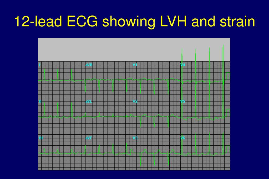
PPT Left Ventricular Hypertrophy PowerPoint Presentation ID537329

Hypertension Left Ventricular Hypertrophy with Strain on ECG Strip

Left Ventricular Hypertrophy LVH with Strain Pattern on ECG YouTube
.jpg)
ECG Interpretation ECG Interpretation Review 51 (Chamber Enlargement
Left ventricular hypertrophy (LVH) with strain pattern

How to differentiate LV strain pattern from primary LV ischemia ? Dr

The ECG in left ventricular hypertrophy (LVH) criteria and
PPT ECG PRACTICAL APPROACH PowerPoint Presentation, free download

ECG in left ventricular hypertrophy (LVH) criteria and implications

ECG showing LVH (left ventricular hypertrophy patternSV 1 + RV 5 = 46
It Can Also Cause Changes To The Heart’s Conduction System That Make It Beat Irregularly (Arrhythmia).
It Can Result In A Lack Of Oxygen To The Heart Muscle.
Note That The Y ‐Axis Scales Vary By Outcome.
Web This Multiethnic Study Of Adults Without Past Cardiovascular Disease Showed That Ecg Strain Is Associated With A Higher Risk For All‐Cause Death, Incident Heart Failure, Myocardial Infarction, And Incident Cardiovascular Disease Independent Of Ecg Left Ventricular (Lv) Hypertrophy Measured By Qrs.
Related Post: